Internation Journal of Myriapodology 5: 49–53, doi: 10.3897/ijm.5.1995
A callipodidan cocoon (Diplopoda, Callipodida, Schizopetalidae)
Henrik Enghoff1, Nesrine Akkari1
1 Natural History Museum of Denmark, University of Copenhagen, Universitetsparken15, DK-2100, København Ø, Denmark
Abstract
A cocoon produced by a juvenile Prolysiopetalum scabratum (L. Koch, 1867) is described and illustrated. SEM pictures of the threads are provided and previous literature on spinning in callipodidan millipedes is reviewed.
Keywordsmillipede, spinning, spinneret, moulting, SEM images
Introduction
The millipede order Callipodida belongs to the superorder Nematophora together with two further orders: Chordeumatida and Stemmiulida. The Nematophora are, i.a., characterized by having two or more spinnerets on the telson (Enghoff 1984). Telsonian spinnerets have also been reported from species of the order Polydesmida, superorder Merocheta, and possibly the very poorly known order Siphoniulida (Shear 2008).
The actual function of the spinnerets has been described only in a handful of cases, most of which concern species of Chordeumatida. Verhoeff (1926-32: 1056-1064) described webs produced by species of Chordeumatida in great detail and summarised most of the earlier literature on the subject; see also Cook (1897), Youngsteadt (2008) and especially Main (1931) who provided a very nice account of moulting nests and egg nests spun by Nanogona (=Polymicrodon) polydesmoides (Leach, 1814), fam. Craspedosomatidae.
In Polydesmida, the function of the spinnerets has been described for two species of Pyrgodesmidae, Poratia obliterata (Kraus, 1952) and Poratia digitata (Porat, 1889), by Adis et al. (2000). Juveniles of both species weave silken threads of different consistence and thickness to cover the soil and the ceiling of the moulting chamber built of decaying wood, soil material, etc (Adis et al. 2000). The spinnerets of Poratia spp. seem to be non-functional in adults. Nothing is known of the function of spinnerets in Stemmiulida and Siphoniulida.
For Callipodida, the earliest report on spinning activity dates back to Fanzago (1874) who described a cocoon produced by “Lysiopetalum carinatum Brandt, 1840”, a species of uncertain identity, certainly not Acanthopetalum carinatum (Brandt, 1840) which is a purely Balkan species (Kime and Enghoff 2011). This cocoon, of variable diameter, is uniformly ochre yellow, ovoid in shape, and shelters the moulting animal, coiling inside. In his description, Fanzago (1874) mentioned two distinct layers forming the cocoon, the peripheral one being more compact and devoid of any aperture. According to Fanzago (1874) the cocoon consists of a great number of continuous and intertwined silk filaments, each “1/269 mm” in diameter. The compact structure of the moulting chamber suggests that it is due to a secretion which could be produced by the spinnerets (Shear 2008; Silvestri 1903; Fanzago 1874).
Since 1874, the only published observations on callipodidan spinning are due to Nguyen Duy-Jacquemin (1979) who observed the moulting of the first two juvenile stadia of Callipus foetidissimus Savi, 1819, fam. Callipodidae. The first juvenile stadium of Callipus foetidissimus produces a thread for closing the opening of its moulting chamber. The second stadium closes the cavity which it occupies with claylike pellets originating from its anus and then weaves a lodge of threads inside. A ‘silken' molting chamber was also noticed for Abacion texense (Loomis, 1937), fam. Abacionidae, although its construction was not observed (N. Youngsteadt, personal communication). The density of threads produced by both these species is low, and there is no resemblance of a compact cocoon like the one described by Fanzago (1874).
Silvestri (1903) investigated callipodidan spinnerets histologically and found the four multicellular glands serving the spinnerets to be essentially the same as in the Chordeumatida, but in adult callipodidans the glands were described as “atrophied.”
Material and methods
Living material of Prolysiopetalum scabratum (L. Koch, 1867) was kept for observation in a terrarium at room temperature in the Natural History Museum of Denmark. Observations and measurements of the cocoon and silken threads were made using a stereomicroscope Leica Wild M10. The photograph was processed with a Leica DFC 420 digital camera mounted on a Leica MZ16A stereomicroscope, and obtained after a final stacking made with Helicon Focus 4.60.2 Pro software program. SEM pictures were made with a JEOL JSM-6335F scanning electron microscope and processed with Adobe photoshop CS5.
Results
Four juveniles of Prolysiopetalum scabratum (L. Koch, 1867) (Schizopetalidae) were collected in GREECE, Athens, S slope of Akropolis, 37°58'15"N, 23°43'40"E, 16.i.2011 by HE.Although the postembryonic stadium was not determined it would have been at least stadium V (in analogy with other callipodidans, cf. Enghoff et al. 1993); the body length was ca. 20 mm.
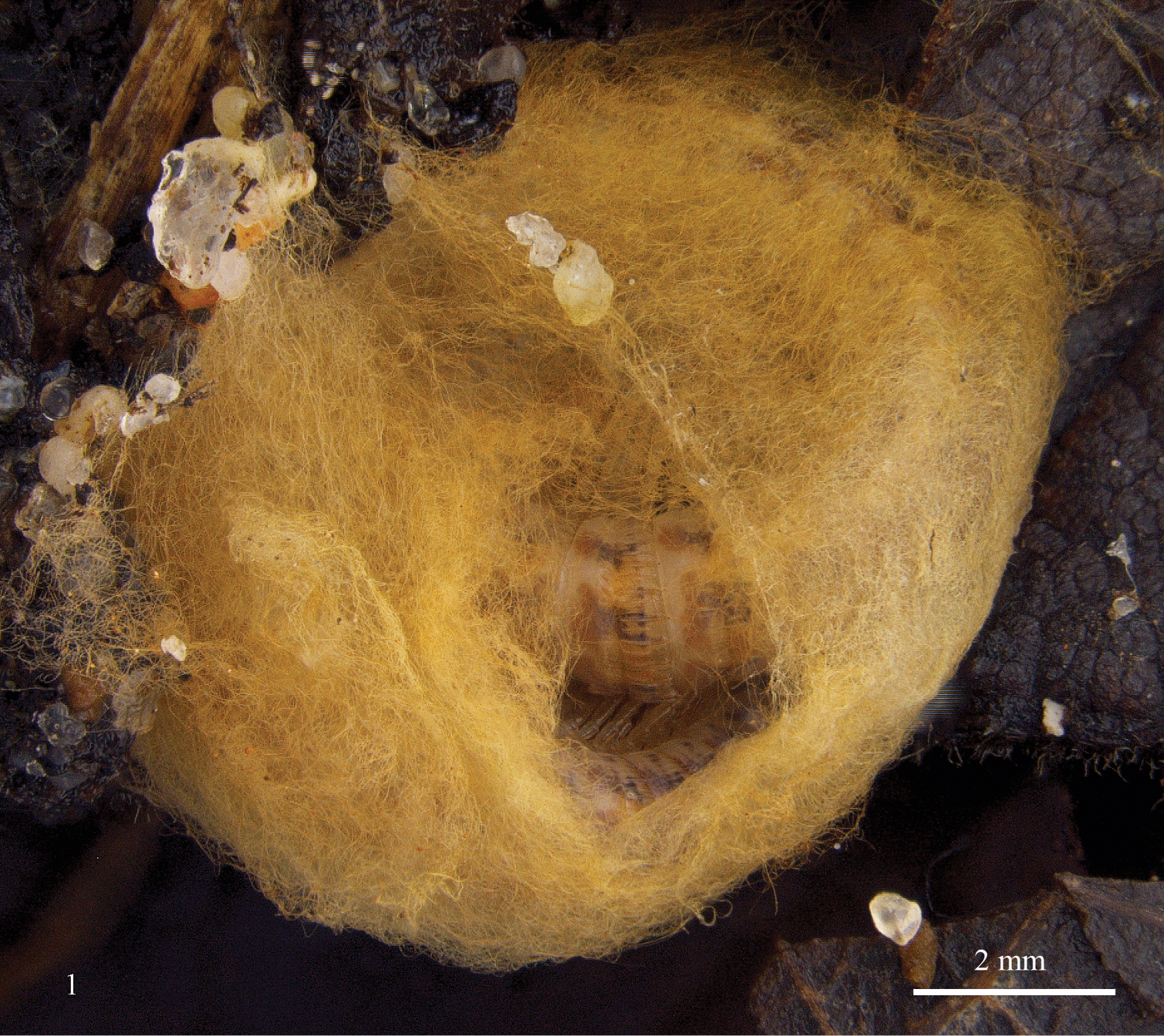
On 4.iv.2011, one of the specimens was found lying inside a densely woven cocoon (Fig. 1). The cocoon is ochre yellow, subcircular, measures approximately 9 mm in diameter by 4 mm in height and closely fits the description given by Fanzago (1874) except for the diameter of the threads which we found to vary between ca. 0.001 and 0.0025 mm, compared to Fanzago's “1/269 mm” (= 0.0037 mm).
Figure 1.
Cocoon of Prolysiopetalum scabratum in terrarium.
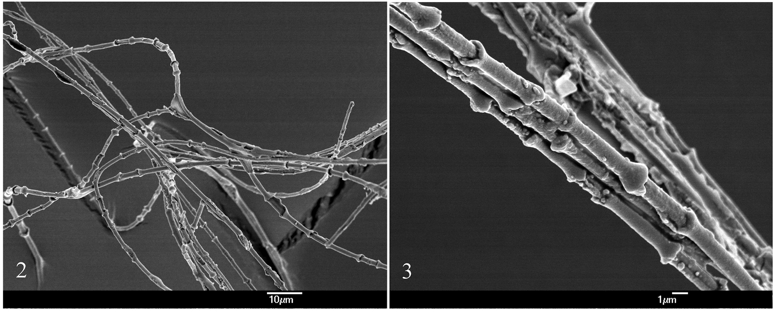
The threads bear tiny subspherical nodules, as is also the case for chordeumatidan threads (Verhoeff 1926-32); the distance between the nodules is approximately 0.005 mm. In the scanning electron microscope the nodules are seen to represent the swollen ends of tiny cylinders which seem to surround a central, thinner thread (Figs 2, 3). The aperture in the cocoon (seen in Fig. 1) was made by us during our examination.
A few weeks later the moulted specimen (still juvenile/subadult) had emerged from the cocoon. It was left alive but perished after some weeks. Two of the other juveniles were found dead in partly completed cocoons.
This observation constitutes the first report of a callipodidan cocoon since 1874 and is presented here in the hope that others will look for similar cocoons and study their formation and structure in more detail than we have been able to do.
Figures 2–3.
Scanning electron micrographs of threads from the cocoon of Prolysiopetalum scabratum 2general view 3 close-up of three parallel threads.
Acknowledgements
We are grateful to Monique Nguyen Duy-Jacquemin (Paris, France), Norman Youngsteadt (Springfield, Missouri, USA) and Alessandro Minelli (Padova, Italy) for information and help with obtaining a copy of Fanzago's paper.
ReferencesAdis J, Hansen B, Wilck L, Adis I, Hirschel K (2000) A spinning apparatus documented in Polydesmida for the first time. In: Wytwer J, Golovatch SI (Eds). Progress in studies on Myriapoda and Onychophora. Fragmenta faunistica, Supplement 43: 139-149.
Cook OF (1897) A spinning diplopod. Brandtia 9: 41-42.
Enghoff H, Dohle W, Blower JG (1993) Anamorphosis in millipedes (Diplopoda) – the present state of knowledge with some developmental and phylogenetic considerations. Zoological Journal of the Linnean Society 109: 103-234.
doi: 10.1111/j.1096-3642.1993.tb00305.x
Kime RD, Enghoff H (2011) Atlas of European millipedes (Class Diplopoda), volume 1, orders Polyxenida, Glomerida, Platydesmida, Siphonocryptidae, Polyzoniida, Callipodida, Polydesmida. Fauna Europaea Evertebrata 3, Pensoft, Sofia-Moscow, 282 pp.
Main H (1931) A millipede's tent. The Essex Naturalist 23: 203– 206.
Nguyen Duy-Jacquemin M (1979) Contribution à l'étude du développement postembryonnaire de
Callipus foetidissimus Savi, 1819 (Myriapode, Diplopode). II. De l'éclosion au stade III. Bulletin du Muséum National d'histoire naturelle de Paris, 4e série, Section A, Zoologie 1 (1): 79–93.
Shear WA (2008) Spinnerets in the millipede order Polydesmida, and the phylogenetic significance of spinnerets in millipeds (Diplopoda). International Journal of Myriapodology 2: 123-146.
doi: 10.1163/187525408X395904
Silvestri F (1903) Classis Diplopoda. Vol. 1a. Anatome: Pars I, Segmenta, Tegumentum, Musculi. In: Berlese A. Acari, Myriapoda et Scorpiones huscque in Italia reperta 346: 1–272. Portici.
Verhoeff KW (1926–1932) Gliederfüssler: Arthropoda, Klasse Diplopoda. In HG Bronn: Bronn's Klassen und Ordnungen des Tier-Reichs, 5, II Abteilung, 2 Teil, 7–13 Lieferung, iii–vi: 1073–2084.
Youngsteadt NW (2008) Laboratory observations on the behavior of two troglobitic millipede species in the genus
Causeyella (Chordeumatida:Trichopetalidae) from the southern Ozarks. Transactions of the Kansas Academy of Science 111: 136-140.
doi: 10.1660/0022-8443(2008)111[136:LOOTBO]2.0.CO;2